
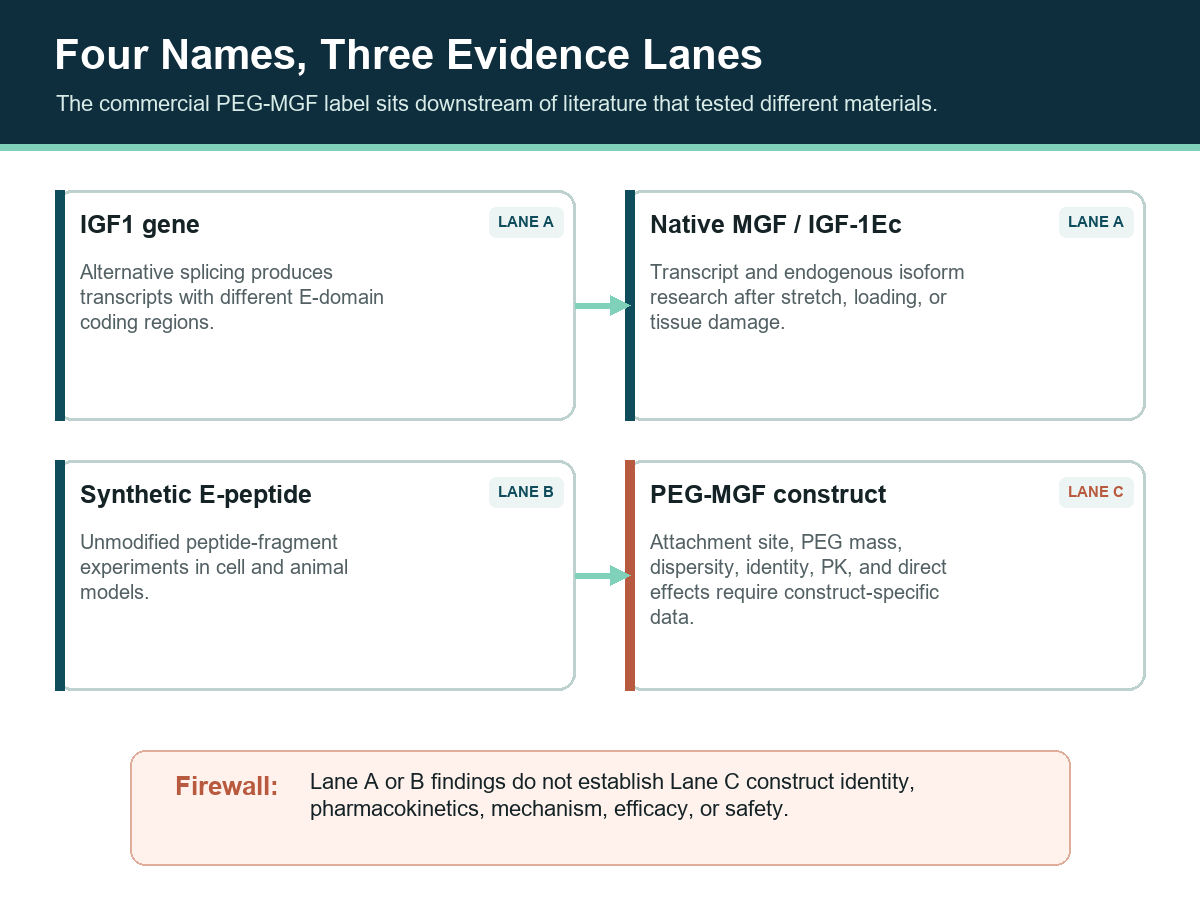
PEG-MGF is a supplier-defined PEGylated research construct commonly described as a polyethylene-glycol-linked form of the Mechano Growth Factor E-peptide. Native MGF and PEG-MGF are not the same compound. The PEG-MGF label does not, by itself, define the peptide sequence, PEG mass or distribution, attachment site, molecular formula, or pharmacokinetic profile. More importantly, the selected peer-reviewed literature largely studies native IGF1 splice-variant biology or an unmodified synthetic MGF E-peptide—not the commercial PEG-MGF construct.
This guide separates those materials into evidence lanes before reviewing IGF1 splicing, mechanically responsive expression, E-peptide experiments, conflicting replication, and minimum construct-characterization controls. This evidence boundary prevents adjacent MGF biology from being presented as direct evidence for PEG-MGF. The guide does not infer PEG-MGF effects from native MGF, provide a personal-use protocol, or treat supplier half-life claims as peer-reviewed findings.
- PEG-MGF is a supplier-defined PEGylated research construct; no standardized sequence, PEG mass, attachment site, or single molecular formula can be assumed from the name alone.
- The molecular weight on the current-lot analytical record is 2,867.20 Da expected and 2,867.36 Da found. The previously published specification of roughly 24,000–26,000 g/mol is withdrawn as an error and is not restored anywhere in this guide.
- A 2,867 Da species is the mass scale of an unmodified 24-residue peptide, not of that peptide carrying the multi-kilodalton PEG chain the withdrawn specification implied; which material the analytical value describes is an open first-party question.
- The selected peer-reviewed corpus primarily studies IGF1 splice variants, native MGF/IGF-1Ec expression, or an unmodified synthetic MGF E-peptide.
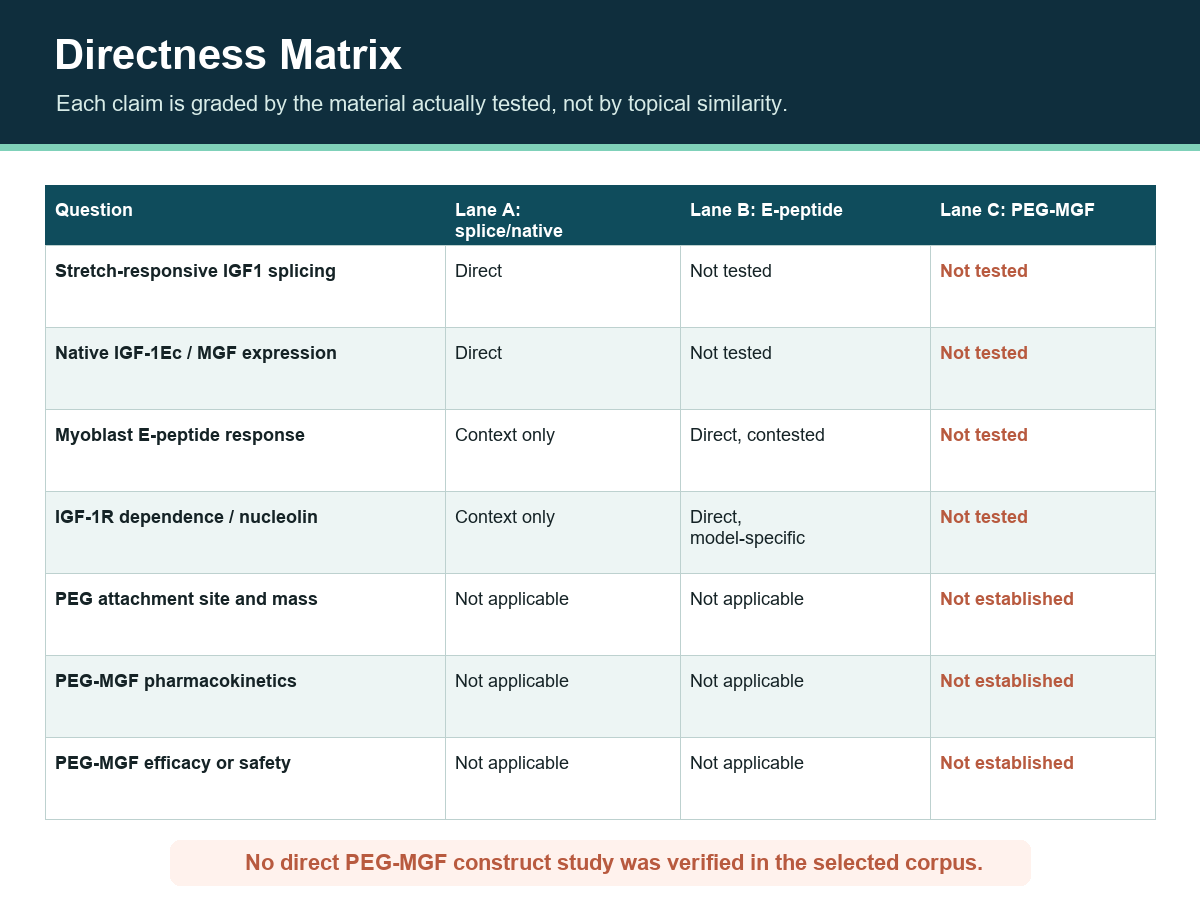
- No direct peer-reviewed study of the commercial PEG-MGF construct was identified and verified in the selected corpus.
- Native MGF expression after stretch or tissue damage does not establish PEG-MGF pharmacokinetics, mechanism, efficacy, or safety.
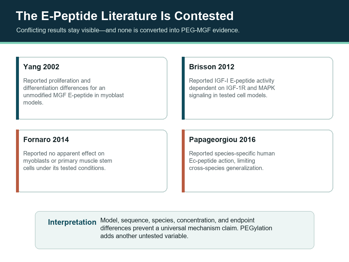
- Unmodified E-peptide findings are contested: the selected literature includes positive, IGF-1R-dependent, null, and species-specific results.
- Claims such as a fixed 48–72-hour half-life remain excluded unless a construct-specific peer-reviewed source and matching material characterization are available.
- Neither PEG-MGF nor native mechano growth factor nor any synthetic MGF E-peptide holds regulatory approval anywhere globally.
What PEG-MGF Means—and What the Name Leaves Unknown
The name combines two ideas: an MGF-related peptide fragment and a polyethylene glycol conjugate. A reproducible chemical identity requires more. At minimum, the exact peptide sequence, terminal modifications, PEG chemistry, nominal or distributed PEG mass, conjugation site, counterion, and analytical confirmation must be stated. Different choices can create materially different conjugates while retaining the same informal label.
The underlying biological molecule is better defined than the conjugate. Mechano Growth Factor is a name given to a particular alternatively spliced product of the IGF1 gene, and the fragment of interest within it is the unique C-terminal E-domain (Ec) peptide of 24 residues. That 24-residue boundary is not an approximation invented by suppliers: Philippou and colleagues raised a rabbit polyclonal antiserum against a synthetic peptide corresponding to the last 24 amino acids of the human MGF E domain and showed that it recognized the E-peptide but not the mature IGF-1 core shared by every isoform, binding MGF in human skeletal muscle and in rat cardiomyocytes and vascular smooth-muscle cells (Philippou et al., 2008). That characterization paper reports no quantitative endpoint, which is expected for an antibody-specificity study, and it is why the field means this particular fragment when it says “MGF peptide.”
A lyophilized presentation does not resolve the remaining identity fields; physical form is not molecular characterization. Neither is a purity percentage. A specification such as ≥99% by HPLC can state an acceptance threshold for the assigned peak’s share of integrated detector signal under a specified method. It does not report whether that peak is the intended conjugate, the free peptide, or a mixture of both; it does not report PEG mass distribution; and it does not report attachment site. For a polymer-peptide hybrid, the gap is closed by orthogonal characterization—mass spectrometry with a stated deconvolution method, alongside a separation that resolves free PEG from free peptide.
Reagent Identity and the Molecular-Mass Correction
A reagent is identified by fields another laboratory can check: sequence, formula, mass, a registry number, and an analytical record tying a specific lot to those values. For this construct, some of those fields are documented, one of them was documented incorrectly, and several do not exist at all. Publishing the table with its gaps visible is more useful than publishing a tidy specification that cannot be reproduced.
The withdrawn mass specification
Mass is the field that changed. An earlier version of this guide carried a specification of approximately 24,000–26,000 g/mol, reasoning that a PEG chain would dominate the conjugate mass. That figure is withdrawn and is not restored. The current-lot analytical record reports an expected mass of 2,867.20 Da and a found mass of 2,867.36 Da. The 0.16 Da difference between expected and found sits inside ordinary mass-spectrometry tolerance for a peptide of this size; the difference between either value and the withdrawn specification does not—2,867 Da is roughly one ninth of 26,000 g/mol.
A species of 2,867 Da is the mass scale of an unmodified 24-residue peptide. It is not the mass of that peptide carrying the multi-kilodalton PEG chain the withdrawn specification implied. Two readings remain open on the record available for this review: the analytical value may describe unconjugated peptide, or it may describe a fragment, an ion or adduct, or a deconvoluted signal rather than the intact conjugate. Distinguishing them requires a frozen construct definition—sequence, terminal modifications, PEG chemistry, nominal and distributed PEG mass, attachment site, counterion, and an expected intact-mass range—which is the subject of an open first-party identity review. Until that review closes, this guide states the analytical figure, states plainly that it conflicts with the withdrawn specification, and declines to assert which material the number describes.
| Field | Documented value | Basis and limits |
|---|---|---|
| Parent gene product | Human IGF-1Ec (mechano growth factor), an alternatively spliced IGF1 transcript | Isoform nomenclature from the IGF-1Ea / IGF-1Eb / IGF-1Ec review literature (PMID 17354613) |
| Bioactive fragment | C-terminal E-domain (Ec) peptide, 24 residues | The 24 most C-terminal residues are the entity synthesized and immunologically characterized in the primary record (PMID 18396778; PMID 20130113) |
| Amino-acid sequence | Not published here | No sequence appears in the cached primary records or in the first-party documentation available for this pass. A verified sequence is required for reagent identity; its absence is recorded as an open blocker rather than filled from recall |
| Molecular weight | 2,867.20 Da expected; 2,867.36 Da found (current-lot analytical record) | Supersedes the withdrawn ~24,000–26,000 g/mol specification. Consistent with an unmodified 24-residue peptide rather than with a multi-kilodalton PEG conjugate; which material the value describes is unresolved |
| Molecular formula | Not assignable as one discrete value. PEG component: ethylene-oxide repeat unit C2H4O, general polymer formula H(OCH2CH2)nOH | A polymer-peptide hybrid with chain-length dispersity has no single formula by definition. No formula for the 24-residue peptide itself is documented in the records available for this pass |
| CAS Registry Number | Polyethylene glycol (polymer component): 25322-68-3 | No CAS number is assigned to the conjugate or to the 24-residue MGF E-peptide. The number above identifies the polymer class only and must not be read as a conjugate registration |
| Physical form | Lyophilized powder | Physical form is a handling attribute, not molecular characterization |
| Purity specification | Read the applicable PEG-MGF report for its assigned species, conjugate characterization, methods and results | Reports main-peak fraction under one method; does not establish conjugation, PEG mass distribution, or attachment site |
| Regulatory status | No FDA, EMA, NMPA, MHRA, PMDA, TGA or Health Canada approval; no approved indication anywhere globally | Applies to PEG-MGF, to native mechano growth factor, and to every synthetic MGF E-peptide described in this guide |
Two of those rows are deliberately empty. An identity table that invents a sequence or a conjugate formula to look complete is worse than one that names the gap, because a fabricated identity field propagates into every downstream document that quotes it.
Native MGF and the IGF1 Splicing Lane
MGF emerged from research on alternative IGF1 gene splicing. A single IGF1 gene produces multiple messenger-RNA isoforms that differ not in their mature IGF-1 core but in their C-terminal extension peptides, the E-domains. Philippou and colleagues set out the vocabulary this article uses—IGF-1Ea, IGF-1Eb, and, in human skeletal muscle, IGF-1Ec—and framed the proposal that proliferation and differentiation are regulated by differential expression of those isoforms after mechanical overload or damage (Philippou et al., 2007). That paper is a narrative review and reports no quantitative finding of its own; it is cited here for nomenclature and for the interpretive frame, not for an effect size.
Cloning the stretch-induced isoform
In rabbit skeletal muscle driven into rapid hypertrophy by active stretch, Yang et al. (1996) cloned and characterized a mechanically responsive IGF-1 isoform. Northern hybridization used a 280-base-pair probe spanning exons 3 and 4 of the IGF1 gene, and two IGF-1 transcripts were strongly expressed in stretched muscle. Resting muscle yielded a single cloned isoform, IGF-1Ea. Stretched, hypertrophying muscle additionally expressed a variant whose E-domain carried a 52-base-pair insert, shifting the reading frame and producing a different carboxy-terminal precursor. With isoform-specific primers, appreciable levels of that variant were detected in rabbit and human skeletal muscle after 2 hours to 6 days of altered physical activity, and were near-absent in control muscle not subjected to stretch.
Which mechanical signal induces the switch
Follow-up work separated the mechanical variables (McKoy et al., 1999). In rabbit extensor digitorum longus muscle, stretch imposed by plaster-cast immobilization in plantar flexion markedly upregulated both IGF-1 mRNA forms within 4 days. Stretch combined with 10 Hz electrical stimulation produced a still larger increase in both transcripts. Electrical stimulation without stretch produced no significant increase over sham-operated controls. Beta-actin expression rose markedly in the stretched and stretched-plus-stimulated muscles, which the authors read as an indication that IGF-1 production may initially be a response to local damage. Type 2X myosin heavy-chain expression fell with stimulation, but less so with stretch alone, suggesting fibre-type conversion runs alongside rather than under IGF-1 control.
These papers are foundational for the “mechano” name, but they studied gene expression and endogenous isoform biology in rabbit muscle. They did not test a PEGylated peptide product. The distinction remains essential when the literature moves from an RNA transcript or endogenous propeptide to an isolated synthetic E-domain fragment.
The transient pulse after damage
Rodent damage models supplied another association. Hill et al. (2003) measured the time course in rat tibialis anterior muscle after two kinds of injury: mechanical damage from electrical stimulation of the stretched muscle, and chemical damage from bupivacaine injection. The autocrine splice variant was expressed rapidly and then declined within a few days, while systemic IGF-1Ea rose more slowly, its increase commensurate with the rate of decline in the autocrine variant. Satellite-cell activation markers M-cadherin and MyoD, including M-cadherin protein, peaked before the IGF-1Ea transcript. The abstract reports no quantitative result for those time courses; the ordering itself is what the model rests on.
Goldspink assembled that ordering into a two-phase account in which mechanical signals shift IGF-1 gene splicing toward the local variant, which “kick starts” hypertrophy and local tissue repair before the systemic isoform supplies mature IGF-1 for protein synthesis (Goldspink, 2005). That review reports no quantitative endpoint either, and it is the origin of the widely repeated claim that impaired expression of the local variant accompanies age-related and disease-related loss of muscle mass. The study supports a temporal relationship in the models tested; it does not establish that a PEG-MGF reagent reproduces the same signal.
Age and the blunted loading response
Owino and colleagues overloaded rat plantaris and soleus by synergist ablation (Owino et al., 2001). Young rats responded with marked hypertrophy and satellite-cell activation indexed by MyoD expression; older muscle did not. The changes associated with that failure were considerably lower expression of the local splice variant and a failure to upregulate IGF-1 receptor and MyoD mRNA. The abstract reports no quantitative finding for the magnitude of those differences, so no effect size is asserted here.
The human counterpart is far more specific. Hameed et al. (2003) studied eight young men aged 25–36 years and seven elderly men aged 70–82 years. Each completed 10 sets of six repetitions of single-leg knee-extensor exercise at 80% of one-repetition maximum, with quadriceps biopsies taken from the exercised and the control leg 2.5 hours after the bout. At rest, transcript levels of the mechanically responsive isoform were approximately 100-fold lower than IGF-1Ea levels, with no difference between the two age groups at rest. High-resistance exercise significantly increased that transcript in the young men but not in the elderly men, while IGF-1Ea mRNA did not change with exercise in either group. Resting MyoD mRNA was greater in the older subjects (P < 0.05), and the older group carried a lower percentage of type II myosin heavy-chain isoforms, though muscle composition showed no association with the isoform response. This is a human observation of endogenous gene splicing under load. It is not an intervention with any peptide, PEGylated or otherwise.
Three Evidence Lanes
A source can be relevant without being direct. The cleanest synthesis divides the corpus into three lanes. Lane A covers IGF1 transcripts, native isoforms, and mechanically responsive expression. Lane B covers experiments using a synthetic, unmodified MGF E-peptide or related E-peptide. Lane C would require explicit testing of a characterized PEG-MGF conjugate.
| Lane | Material studied | What it can support | What it cannot support |
|---|---|---|---|
| A: splice/native | IGF1 transcripts, endogenous isoforms, tissue expression | Splicing, expression timing, model-specific biological association | Synthetic E-peptide identity or PEG-MGF properties |
| B: unmodified E-peptide | Defined synthetic E-peptide without verified PEG conjugation | That peptide’s result in the stated cell/animal model | PEG-MGF PK, mechanism, efficacy, or safety |
| C: PEG-MGF | Characterized PEG-MGF construct | Only the construct, model, endpoint, and conditions directly tested | Universal claims across differently specified supplier constructs |
No direct PEG-MGF evidence was identified and verified in the selected peer-reviewed corpus as of this review. That is not proof that no such paper exists anywhere; it is a transparent limit of the source set used for this article. The correct editorial response is to leave Lane C unfilled rather than converting adjacent MGF literature into construct evidence.
What the Unmodified E-Peptide Studies Actually Tested
One frequently cited Lane B paper compared an MGF E-peptide with mature IGF-I in myoblast systems. Yang and Goldspink (2002) reported that the distinct E domain inhibited terminal differentiation while increasing myoblast proliferation—the opposite division of labour from mature IGF-1—and that blocking the IGF-1 receptor with a specific antibody did not abolish the E-domain effect, which they read as evidence of a different receptor. The abstract reports no quantitative result for that difference, and the record carries a 2006 erratum (FEBS Lett 580(10):2530), so the corrected version is the one to read before quoting anything from it. The material was an unmodified synthetic E-peptide, not a characterized PEG-MGF conjugate.
The human primary-cell extension
Two later studies moved the design into human muscle cultures. Ates et al. (2007) used primary cultures from healthy subjects and from muscular-dystrophy and ALS patients. Initial numbers of desmin-expressing mononucleated progenitor cells were lower in diseased muscle. The E-domain peptide of IGF-1Ec significantly increased progenitor-cell numbers in healthy and in diseased cultures, while IGF-1 enhanced myogenic differentiation and the E-peptide blocked that pathway—the stated mechanism for an enlarged progenitor pool. That abstract reports no quantitative endpoint for the increase.
Kandalla et al. (2011) applied the 24-amino-acid E peptide to primary human muscle cultures from donors of different ages, noting that the human insert is 49 base pairs against 52 in rodents. The peptide significantly increased proliferative life span and delayed senescence in satellite cells isolated from neonatal and young-adult muscle, but not in cells from old-adult muscle, and a significant decrease in the percentage of reserve cells was seen across all cultures. No quantitative endpoint appears in that abstract either. Taken together with the age-related splicing data, this is the strongest form of the sarcopenia argument in the corpus—and it is entirely in-vitro human cell work.
Mixed designs and the receptor question
A later paper combined human exercise-induced muscle-damage expression measurements with C2C12 myoblast experiments using synthetic MGF E-peptide (Philippou et al., 2009). Ten healthy male volunteers underwent exercise-induced muscle damage, with biopsies taken before and at 6 hours, 2, 5 and 16 days after exercise. Expression of the mechanically responsive isoform rose rapidly and transiently, followed by a prolonged increase in IGF-1Ea and IGF-1Eb transcripts (p<0.05), with matching patterns at the protein level. In vitro, the synthetic E-peptide’s proliferative effect on C2C12 cells was not blocked by an anti-IGF-1-receptor neutralizing antibody and it did not phosphorylate Akt. This mixed design contains both a human expression lane and an in-vitro peptide lane. It does not contain a human PEG-MGF intervention.
Mechanism remains model-dependent. Brisson and Barton (2012) synthesized EA, EB and a scrambled control peptide and tested them in murine C2C12 cells. Both E-peptides increased MAPK signalling, and pharmacological IGF-1-receptor inhibition blocked it. Neither peptide directly induced receptor phosphorylation, yet the presence of either increased receptor activation by IGF-1 through greater cell-surface receptor bioavailability. EB increased myoblast proliferation and migration, effects abolished by MAPK or IGF-1-receptor blockade, while EA delayed differentiation. That abstract states no quantitative outcome; its conclusion is directional, and it is the direct opposite of the distinct-receptor proposal—the E-peptides are described as having little independent activity, acting instead by modulating IGF-1 signalling. In neuronal research, Podratz et al. (2020) identified nucleolin interaction in a cisplatin-neurotoxicity model, in which cisplatin induces peripheral neuropathy in 30–40% of treated patients. Different cell types, peptide preparations, and endpoints prevent these findings from becoming one universal pathway—and neither paper establishes PEG-MGF behavior.
Why the E-Peptide Evidence Remains Contested
The selected corpus does not support a one-directional summary. Fornaro et al. (2014) reported no apparent effect of the tested mechano-growth-factor peptide on myoblasts or primary muscle stem cells under its experimental conditions. The replication was attempted independently at two pharmaceutical companies. Concentrations of peptide up to 500 ng/ml failed to increase proliferation of C2C12 cells or of primary human skeletal muscle myoblasts, while mature IGF-1 and full-length IGF-1Eb produced a proliferative response in the same cells. The peptide also failed to inhibit differentiation of myoblasts into myotubes, failed to alter proliferation or differentiation in primary mouse skeletal muscle stem cells, and—as either the native or a stabilized peptide—failed to activate p-ERK in cardiac myocytes, an effect separately documented in the earlier literature. This directly conflicts with broader claims that an MGF E-peptide consistently drives myoblast or satellite-cell responses.
Species adds another boundary. Papageorgiou et al. (2016) characterized the human Ec peptide as having a species-specific mode of action. Human Ec behaved as a progression rather than a competence growth factor in PC-3 prostate-cancer cells, activating ERK1/2 without affecting Akt phosphorylation. A narrow 5–50 nM range stimulated PC-3 growth in medium supplemented with 10% fetal bovine serum; the same peptide did nothing in 0.5% serum, and nothing in mouse C2C12 myoblasts under any culture condition. Its activity was blocked by a neutralizing anti-human-IGF-1Ec antibody but not by an anti-IGF-1-receptor antibody. Synthetic mouse Ec was inactive in human PC-3 cells yet significantly stimulated mouse C2C12 proliferation, and fragment analysis located the active core in the last four residues of the human peptide’s C-terminal end. A source set that contains positive, null, receptor-dependent, and species-specific findings requires a conditional synthesis, not a marketing conclusion.
A critical minireview by Matheny et al. (2010) is useful for understanding the interpretive problems surrounding MGF as a putative IGF-I gene product involved in repair and regeneration. It records one caveat that outranks the rest: a synthetically manufactured peptide corresponding to the 24 most C-terminal residues has been shown to promote cellular proliferation and survival, yet no analogous peptide product of the Igf1 gene has been identified in or isolated from cultured cells, their conditioned medium, or animal tissues or biological fluids. As a review it reports no quantitative endpoint of its own; it organizes the controversy and supplies no direct PEG-MGF evidence.
Findings Outside Skeletal Muscle—Still Lane A and Lane B
The E-peptide literature did not stay in muscle. Brain-ischemia, motoneuron-disease, neurogenesis, myocardial-infarction and osteoblast models each produced reported effects, and each used a synthetic unmodified peptide, a genetic overexpression construct, or an expression plasmid—never a characterized PEGylated conjugate. These are the studies most often paraphrased on supplier pages as though they described PEG-MGF, which is why they are itemized below with their species, model and readout intact.
Central nervous system models
Dluzniewska and colleagues gave the synthetic C-terminal peptide in a gerbil model of transient brain ischemia and reported very significant protection of vulnerable neurons, with ischemia also raising endogenous expression in ischemia-resistant hippocampal neurons (Dluzniewska et al., 2005). In organotypic hippocampal culture the synthetic peptide was as potent as full-length IGF-1 and its effect lasted significantly longer; the two were additive, and the neuroprotective action was independent of the IGF-1 receptor. Riddoch-Contreras and colleagues delivered an expression plasmid carrying either MGF or IGF-1 cDNA into the hindlimb muscles of SOD1(G93A) mice at 70 days of age, at symptom onset, and reported significant improvement in hindlimb muscle strength with increased motor-unit and motoneuron survival, with significantly more motoneurons surviving in the MGF-treated animals (Riddoch-Contreras et al., 2009). Tang and colleagues took the question into ageing, using transgenic and conditionally inducible overexpression in mice (Tang et al., 2017).
These are encouraging-looking preclinical signals in specific models. They carry no implication for human use, and they are included to map the breadth of the research rather than to assert efficacy.
Cardiac and bone models
Carpenter and colleagues induced infarcts in sheep by microsphere injection and treated animals with 200 nM of mature IGF-1, the E domain, or full-length MGF, reporting improved cardiac function with the E domain and, in a second experiment, 35% less compromised cardiac muscle than controls at eight days post-infarction (Carpenter et al., 2008). Mavrommatis and colleagues supplied the mechanistic complement, reporting rapid cellular uptake without IGF-1-receptor activation, nuclear localization, and inhibition of the intrinsic apoptotic pathway in stressed H9c2 cells, followed by preserved contractility on pressure-volume analysis at 2 weeks post-infarction in mice (Mavrommatis et al., 2013). Outside contractile tissue, Xin and colleagues treated the MC3T3-E1 pre-osteoblast line with 1 nM peptide and reported inhibited differentiation and delayed mineralization through increased ERK activity and decreased Cbfα-1 nuclear translocation (Xin et al., 2012)—the same differentiation-inhibiting direction, in a non-muscle cell type.
The delivery study that is not a PEGylation study
One paper is regularly miscited as evidence for PEGylated MGF, and it deserves its own paragraph. Peña and colleagues built poly(ethylene glycol) dimethacrylate hydrogel microstructures sized like an adult cardiac myocyte, at 100 × 15 × 15 µm with a stiffness of 20 kPa, loaded them with the human E-domain peptide, and injected them intramuscularly after coronary-artery ligation in mice (Peña et al., 2015). Untreated animals showed a significant decline in systolic and diastolic function with pathologic hypertrophy by 2 weeks, decompensating further by 10 weeks. Peptide-eluting microrods decreased mortality, ameliorated the haemodynamic decline and delayed decompensation; empty microrods had limited effect.
Note precisely what that construction is. The polyethylene glycol is a crosslinked hydrogel scaffold surrounding the peptide and releasing it locally. It is not a PEG chain covalently attached to the peptide. The study is a delivery-engineering result about a short-lived fragment, and it says nothing about the pharmacokinetics, receptor access or activity of a conjugate. Treating it as a PEG-MGF study conflates a carrier with a covalent modification.
Additional Reported Findings
The table below indexes the remaining studies behind this article, including the model-specific results discussed in the sections above. Each row names the material tested, the model, the species, the endpoint and the reported result, with the source linked so any figure can be checked against its own abstract. Where an abstract reports no effect size, the row says so rather than supplying one.
| Material | Model | Species | Endpoint | Reported result | Source |
|---|---|---|---|---|---|
| Endogenous MGF splice variant | Synergist-ablation overload of plantaris and soleus, young vs old | Rat | Local splice-variant, IGF-1R and MyoD mRNA | Marked hypertrophy and MyoD-indexed satellite-cell activation in young rats; considerably lower splice-variant expression and failure to upregulate IGF-1R and MyoD in old muscle — no quantitative endpoint reported | PMID 11566187 |
| Endogenous IGF-1 splice variants | 10 × 6 single-leg knee extensions at 80% 1RM; quadriceps biopsy 2.5 h post-exercise | Human (8 men aged 25–36 y; 7 men aged 70–82 y) | MGF and IGF-1Ea mRNA; MyoD; MyHC composition | Resting MGF ~100-fold below IGF-1Ea; MGF mRNA rose significantly in young men only; IGF-1Ea unchanged by exercise in both groups; resting MyoD higher in older men (P < 0.05) | PMID 12562960 |
| Narrative review of mechanical signalling and IGF-1 splicing | Review | Rodent and human literature | Two-phase adaptation model | Local variant proposed to initiate hypertrophy and local repair before the systemic isoform; impaired expression linked to age- and disease-related muscle loss — review reports no quantitative endpoint | PMID 16024511 |
| Narrative review of IGF-1 in skeletal-muscle physiology | Review | Human and rodent literature | Isoform nomenclature and proposed roles | IGF-1Ea, IGF-1Eb and IGF-1Ec share the mature core and differ in E-domain; proliferation/differentiation split proposed — review reports no quantitative finding | PMID 17354613 |
| Rabbit antiserum raised against the last 24 aa of the human E domain | Western blot and immunohistochemistry | Rabbit antiserum; human skeletal and rat cardiac muscle | Epitope specificity | Recognized the E-peptide and not the mature IGF-1 core; bound MGF in human skeletal muscle and in rat cardiomyocytes and vascular smooth-muscle cells — no quantitative endpoint reported | PMID 18396778 |
| Synthetic unmodified E-peptide | Primary muscle cell culture: healthy, muscular-dystrophy and ALS donors | Human, in vitro | Desmin-positive progenitor number; differentiation | Progenitor numbers significantly increased in healthy and diseased cultures; IGF-1 enhanced differentiation while the E-peptide blocked it — no quantitative endpoint reported | PMID 17531227 |
| Synthetic 24-amino-acid E peptide | Primary muscle cultures across donor ages (49-bp human insert; 52-bp rodent) | Human, in vitro | Proliferative life span; senescence; reserve-cell fraction | Proliferative life span significantly increased and senescence delayed in neonatal and young-adult but not old-adult satellite cells; reserve-cell percentage significantly decreased in all cultures — no quantitative endpoint reported | PMID 21354439 |
| Synthetic C-terminal (E-domain) peptide | Transient brain ischemia in vivo; organotypic hippocampal culture in vitro | Gerbil in vivo; hippocampal slice in vitro | Neuronal survival | Very significant protection of vulnerable neurons; in vitro as potent as full-length IGF-1 with a significantly longer-lasting effect, additive with it, and independent of the IGF-1 receptor — no quantitative result reported | PMID 16144956 |
| MGF or IGF-1 cDNA expression plasmid, intramuscular | Delivery to hindlimb muscles at 70 days of age, at symptom onset | SOD1(G93A) mouse | Hindlimb muscle strength; motor-unit and motoneuron survival | Significant improvement in strength and in motor-unit and motoneuron survival with either construct; significantly more motoneurons survived with MGF | PMID 19038252 |
| Transgenic and lac-operon-inducible MGF overexpression | Constitutive from birth; induction at 1, 3 or 12 months with scoring at 24 months | Mouse; SVZ-derived neural stem cells in vitro | BrdU+ cells; olfactory function; neurosphere size and number | More BrdU+ cells in dentate gyrus and subventricular zone with unchanged post-mitotic distribution; more BrdU+ proliferating and mature olfactory-bulb neurons with preserved olfaction when induced at 1 or 3 months, but not at 12 months | PMID 28683812 |
| Synthetic E-domain peptide, 200 nM | Microsphere-induced myocardial infarction; assessed 8 days post-MI | Sheep | Cardiac function; compromised myocardium; cleaved caspase-3 | Cardiac function preserved; 35% less compromised cardiac muscle than controls; cleaved caspase-3 immunostaining absent in treated hearts | PMID 17581790 |
| Synthetic E-domain peptide analog | Sorbitol-stressed H9c2 cells; peptide given at time of MI and assayed 2 weeks post-MI | H9c2 cell line; mouse | Mitochondrial membrane potential; caspase-3; PV-loop function; apoptotic nuclei | Nuclear localization without IGF-1R activation; mitochondrial collapse and caspase-3 activation inhibited; significant preservation of systolic and diastolic function; less pathologic hypertrophy and significantly fewer apoptotic nuclei | PMID 23712705 |
| E-domain peptide eluted from PEG-dimethacrylate hydrogel microrods (100 × 15 × 15 µm, 20 kPa) | Intramuscular injection after coronary-artery ligation; 2 and 10 weeks | Mouse | Mortality; PV-loop haemodynamics; pathologic hypertrophy | Untreated hearts declined by 2 weeks and decompensated by 10 weeks; peptide-eluting microrods decreased mortality, ameliorated the decline and delayed decompensation; empty microrods had limited effect | PMID 25678113 |
| Synthetic MGF-E peptide, 1 nM | MC3T3-E1 pre-osteoblast line ± ERK inhibitor PD98059 | Mouse cell line, in vitro | ALP activity; collagen I; osteopontin; Cbfα-1; mineralization | ALP activity decreased with increased ERK activation; collagen I inhibited and osteopontin enhanced; activated nuclear Cbfα-1 decreased; mineralization delayed — each effect weakened by PD98059 | PMID 22186070 |
One source cited by the previous version of this guide is not carried forward. A 2005 review of impaired IGF-1 gene splicing and MGF expression in muscle wasting (PMID 16125110) carries both an erratum and a republication notice, and the indexed abstract on its record does not correspond to its title. No claim from it can be verified in its current form, so it is dropped rather than restored; the same argument is carried by the 2005 Physiology review already cited above.
What PEGylation Does Not Prove
PEG conjugation is a chemical modification, not an evidence shortcut. Changing hydrodynamic behavior or proteolytic exposure is a plausible design objective, but the magnitude and direction of any change depend on PEG size, distribution, linkage, attachment site, peptide integrity, and model. Without construct-specific characterization and direct measurements, a fixed 48–72-hour half-life is not a verified scientific value.
The rationale usually offered for conjugation is that the native fragment is short-lived. That premise is reasonable and is exactly why delivery engineering was attempted in the cardiac work described above. But a rationale is not a measurement. No half-life for the unmodified 24-residue peptide is stated in the sources reviewed here, and no half-life for any PEG conjugate of it appears in them at all. A number with no measurement behind it should be reported as absent, not rounded into a range.
The same rule applies to mechanism. PEG attachment may alter receptor access, tissue distribution, assay behavior, or biological activity. A positive result for an unmodified E-peptide cannot establish that the PEGylated construct retains, enhances, or prolongs that result. A null unmodified-peptide result also cannot establish that every PEG conjugate is inactive. Lane C needs Lane C experiments.
Research-Design Framework for PEG-MGF
A rigorous design begins with material characterization. The exact peptide and PEG attributes should be recorded before comparing biological results. Analytical identity should distinguish free peptide, free PEG, intended conjugate, and possible mixtures; a generic purity percentage alone cannot establish conjugation identity. Researchers evaluating batch documentation can use the COA-reading guide and HPLC testing explainer as method context while recognizing that a construct may require orthogonal characterization.
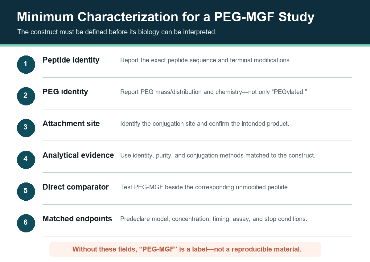
The experimental comparison should include the corresponding unmodified E-peptide, vehicle and assay controls, matched concentrations where analytically defensible, prespecified time points, and endpoints selected before results are known. Any interpretation must remain attached to the measured construct and model. A result from a cell line, rodent tissue, or neuronal system is not automatically a result for human muscle or another supplier’s conjugate.
The literature reviewed above also supplies the comparator conditions worth copying. Serum concentration changed the outcome in the PC-3 work; species changed it in the same paper; receptor blockade changed it in opposite directions across three groups; and donor age changed it in human primary cells. A construct study that omits those axes will produce a result that cannot be placed alongside the existing record.
Research-Material Governance and Regulatory Status
PEG-MGF holds no marketing authorization anywhere in the world. It is not approved by the FDA, the EMA, the NMPA, the MHRA, the PMDA, the TGA or Health Canada; it has no approved indication in any jurisdiction; and no approved formulation of it exists on the record available for this review. The same applies to native mechano growth factor and to every synthetic MGF E-peptide described in this guide: none has regulatory approval anywhere globally. That is a statement about the compound. A supplier-status claim—that a vendor sells research materials—is a different and much weaker assertion, and it must not be read as a substitute for regulatory status.
This guide intentionally provides no product-page handoff. A commercial listing should be connected to the article only after current first-party peptide identity, PEG specification, attachment chemistry, and mass fields are internally consistent and traceable to source documentation. The unresolved mass conflict described earlier is precisely that condition, and it remains open. Until then, product copy cannot be used to fill the Lane C evidence gap.
For adjacent biology, the IGF-1 LR3 research guide covers a different, full IGF-1 analog research lane, while the growth-hormone-axis hub maps broader cluster relationships. These pages do not imply equivalence. The regulatory boundary between a research-grade chemical reagent and an approved pharmaceutical formulation is set out in the research-grade versus pharmaceutical-grade reference; Apex Laboratory supplies research-use-only chemical reagents intended exclusively for in-vitro and preclinical research.
Frequently Asked Questions
What is PEG-MGF?
PEG-MGF is an informal supplier label for a PEG-conjugated MGF-related peptide research construct. The name alone does not define the exact peptide, PEG mass, attachment chemistry, conjugation site, or molecular composition.
What is the molecular weight of PEG-MGF?
The current-lot analytical record reports 2,867.20 Da expected and 2,867.36 Da found. An earlier specification of roughly 24,000–26,000 g/mol is withdrawn as an error. A 2,867 Da species is the mass scale of an unmodified 24-residue peptide rather than a multi-kilodalton conjugate, and which material the analytical value describes has not yet been resolved in the first-party record.
Does PEG-MGF have a CAS number or a molecular formula?
No CAS Registry Number is assigned to the conjugate or to the 24-residue MGF E-peptide. Polyethylene glycol as a polymer class carries CAS 25322-68-3, which identifies the polymer component only. No single molecular formula can describe a polymer-peptide hybrid with chain-length dispersity; the PEG component is built from the ethylene-oxide repeat unit C2H4O.
Is native MGF the same material as PEG-MGF?
No. Native MGF or IGF-1Ec refers to an endogenous splice-variant context. PEG-MGF describes a synthetic PEGylated construct. Evidence from the native lane cannot be assigned directly to the conjugate.
Do the cited E-peptide studies test PEG-MGF?
No selected E-peptide paper in this guide explicitly tests a characterized commercial PEG-MGF construct. Those sources are presented as Lane B background and are labeled accordingly.
Does PEG-MGF have a verified 48–72-hour half-life?
Not in the selected peer-reviewed corpus. A fixed duration requires a construct-specific pharmacokinetic source and a material matching the tested peptide, PEG, linkage, and attachment site.
Does the literature establish one MGF E-peptide mechanism?
No. The selected record includes proposed distinct-receptor effects, IGF-1R-dependent activity, nucleolin interaction in a neuronal model, a null myoblast replication, and species-specific behavior. These findings are model-specific and do not establish PEG-MGF mechanism.
Why is the MGF research called contested rather than emerging?
Because a well-resourced independent replication reported the opposite of the founding result. Two pharmaceutical companies tested peptide concentrations up to 500 ng/ml and found no increase in proliferation of C2C12 cells or primary human myoblasts, while mature IGF-1 was active in the same systems. A field with a documented failed replication at that scale is contested, and describing it as merely early would misstate the evidence.
Is PEG-MGF approved by any regulator?
No. Neither PEG-MGF nor native mechano growth factor nor any synthetic MGF E-peptide holds FDA, EMA, NMPA, MHRA, PMDA, TGA or Health Canada approval, and none has an approved indication anywhere globally.
What evidence would make a PEG-MGF claim direct?
The paper would need to define and analytically characterize the PEG-MGF construct, test it in the named model, measure the stated endpoint, and report enough material detail to determine whether the evaluated reagent matches.
Continue Your Research
- Growth Hormone Axis Research Peptides
- IGF-1 LR3 Research Guide
- How to Read a Peptide Certificate of Analysis
- Research-Grade vs Pharmaceutical-Grade
- Apex Laboratory Research Library
Research Use Disclaimer
This article is provided for educational and research reference purposes only. PEG-MGF and all products sold by Apex Laboratory are intended exclusively for in-vitro laboratory research use and are not for human consumption. Researchers should consult the primary peer-reviewed literature cited throughout this article for detailed methodological protocols, experimental designs, and complete data sets.