
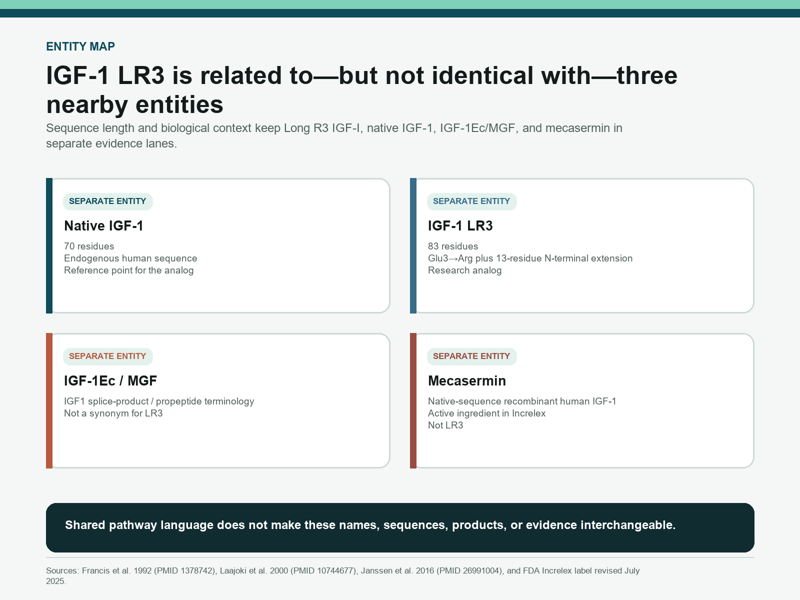
IGF-1 LR3 is an 83-residue analog of human IGF-1 with a Glu3-to-Arg substitution and a 13-residue N-terminal extension. Those changes reduce affinity for insulin-like growth factor binding proteins, making IGFBP context central to its experimental behavior. Direct evidence is largely cell and animal research; native IGF-1, IGF-1Ec/MGF, mecasermin, and general IGF1R findings do not transfer automatically to LR3.
Long R3 IGF-I is easy to describe inaccurately because it sits near several better-known terms. Native IGF-1 explains much of the pathway vocabulary. IGF-binding proteins shape ligand distribution. Mechano growth factor appears in muscle literature. Mecasermin is an approved recombinant human IGF-1 product. None of those facts turns them into the same molecule or the same evidence object as IGF-1 LR3.
- IGF-1 LR3 combines a 13-residue N-terminal extension with an Arg substitution at position 3 of the IGF-I domain, producing an 83-residue analog.
- The design has reduced affinity for IGF-binding proteins, but lower IGFBP binding does not guarantee one potency ranking, half-life, or biological outcome.
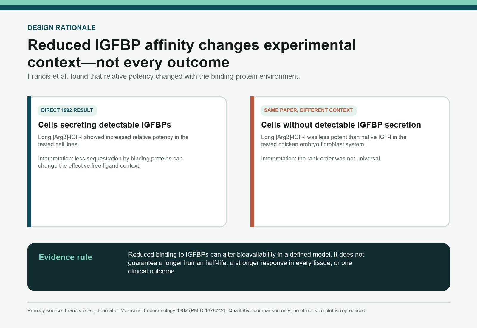
- Francis et al. found that relative potency changed with whether tested cells secreted detectable IGFBPs.
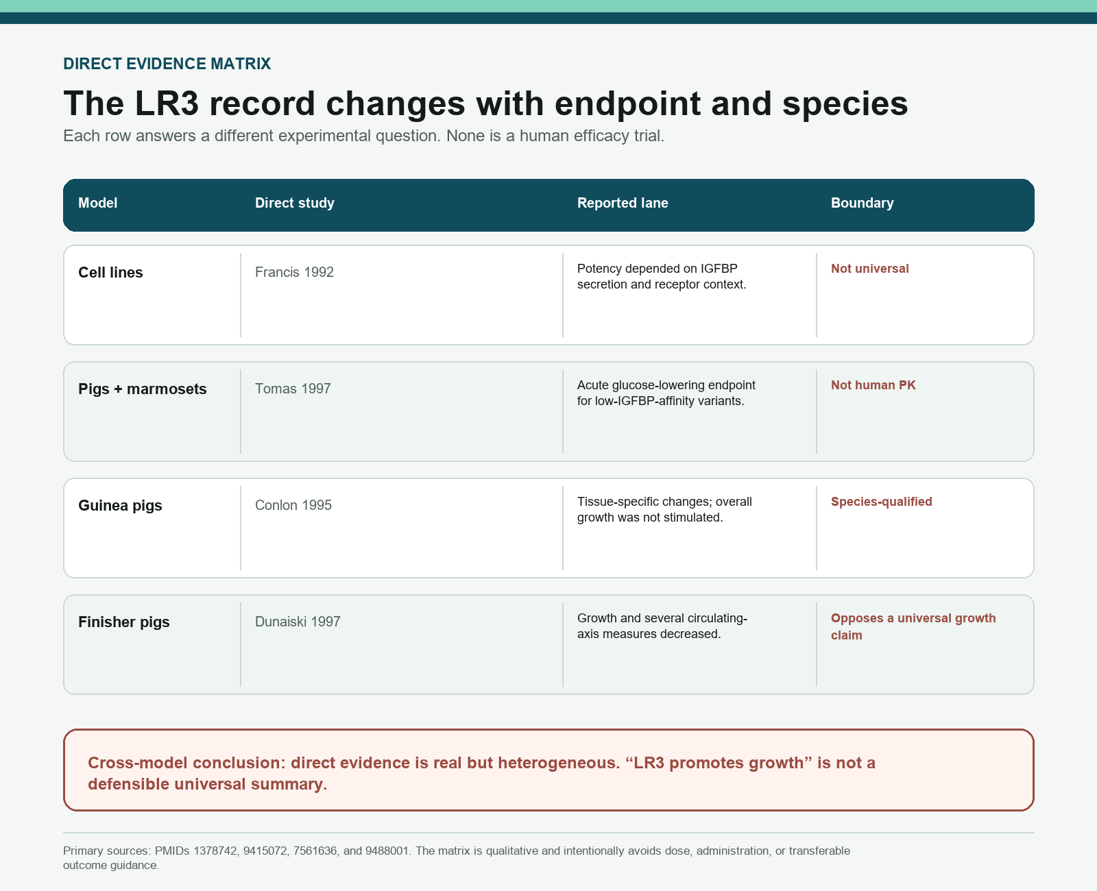
- Direct animal findings are heterogeneous: guinea-pig tissue observations and reduced growth in finisher pigs cannot be compressed into a universal “growth” claim.
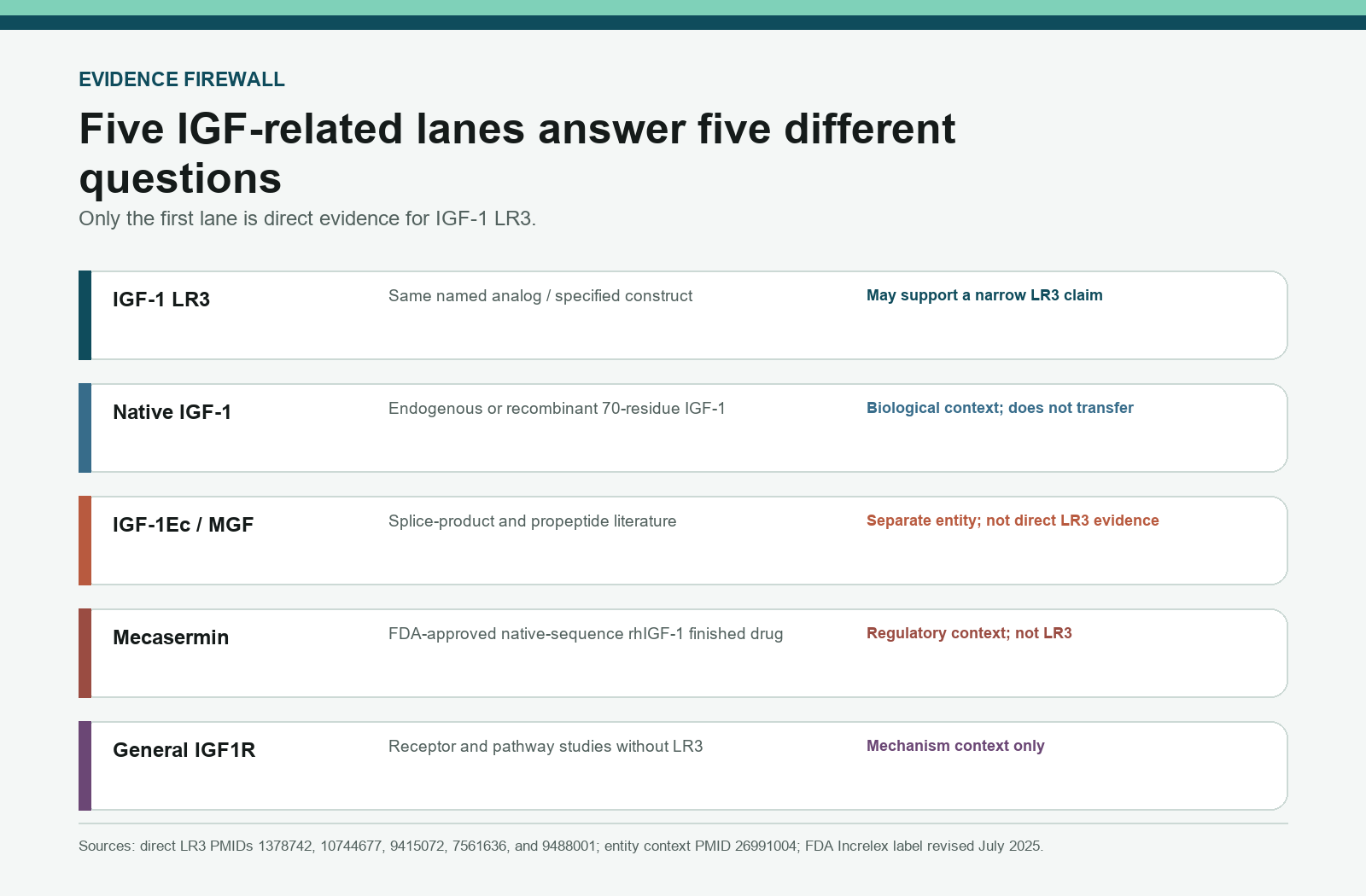
- Native IGF-1, general IGF1R signaling, IGF-1Ec/MGF, and mecasermin evidence are adjacent lanes, not direct LR3 evidence.
- The direct source record does not establish a human LR3 efficacy profile, clinical safety dossier, or universal human pharmacokinetic value.
What Is IGF-1 LR3?
IGF-1 LR3, also written Long R3 IGF-I or Long [Arg3]-IGF-I, is a recombinant analog built around the human IGF-I domain. Its two defining changes are a Glu-to-Arg substitution at position 3 of the IGF-I portion and a 13-residue extension at the N terminus. Human IGF-1 has 70 residues; adding the extension produces the 83-residue LR3 analog.[1][2]
Reagent identity
| Property | Value |
|---|---|
| CAS number | 946870-92-4 (catalog-derived; no PubChem CID for this protein analog) |
| Molecular weight | 9,117.5 Da (≈9.1 kDa), reduced chain |
| Molecular formula | C400H625N111O115S9 reduced, C400H619N111O115S9 with three disulfides (9,111.5 Da); summed from the sequence below, which reproduces the catalog weight |
| N-terminal extension | Met-Phe-Pro-Ala-Met-Pro-Leu-Ser-Ser-Leu-Phe-Val-Asn (13 residues) |
| Sequence | MFPAMPLSSLFVNGPRTLCGAELVDALQFVCGDRGFYFNKPTGYGSSSRRAPQTGIVDECCFRSCDLRRLEMYCAPLKPAKSA (Arg for Glu at position 3) |
| Solution structure | Three α-helices, with the NMR structure determined to a precision of 0.82 ± 0.28 Å backbone RMSD in those helices; N-terminal extension flexible (PMID 10744677) |
| Purity specification | Read the applicable IGF-1 LR3 report for its declared construct, methods, results and acceptance criteria |
The original Francis paper described Long [Arg3]-IGF-I as one of several fusion-protein analogs designed to investigate the relative importance of receptor binding and IGF-binding-protein interactions.[1] That origin matters. LR3 was not designed as a generic synonym for IGF-1; it was designed as an experimental analog whose altered binding-protein interactions could help separate parts of the IGF system.
The name does not establish that every current material is identical to the paper construct. Expression system, folding, disulfide pairing, termini, counterions, aggregation, purity, and lot-specific identity all require separate documentation. An article can define the expected analyte, but only current analytical evidence can describe a particular lot.
What Does the LR3 Design Change?
The Arg3 substitution and N-terminal extension alter the region most involved in interaction with IGF-binding proteins. Solution-NMR work confirmed that the core IGF-I domain remains broadly consistent with IGF-I while identifying N-terminal reorientation and flexibility relevant to lower IGFBP affinity.[2] This is a structural explanation, not a claim that every downstream response must be larger or longer.
“Lower IGFBP affinity” is also more precise than “does not bind IGFBPs.” Relative affinity can vary with the binding protein, assay, and conditions. A reduced interaction can change the fraction of ligand available to receptors, its distribution between compartments, and the timing of an assay readout. It can also remove a form of buffering that native IGF-1 experiences in many systems.
The analog still interacts with the IGF signaling system, but receptor context matters. IGF1R and insulin-receptor isoforms can produce overlapping yet non-identical observations, and a cell line’s receptor abundance changes the result. A pathway diagram built from native IGF-1 cannot substitute for a direct LR3 experiment.
Why Does IGFBP Affinity Matter?
IGF-binding proteins do more than extend circulation time. They can sequester ligand, regulate transport, influence local availability, and change which concentration reaches a receptor during an experiment. The Francis study is especially valuable because it tested IGF-I analogs across cell systems with different binding-protein contexts.[1]
In cell lines that secreted IGFBPs into the medium, Long [Arg3]-IGF-I showed greater relative potency than native IGF-I for the tested endpoints. In chicken embryo fibroblasts without detectable IGFBP secretion, Long [Arg3]-IGF-I was less potent than native IGF-I. The same paper therefore contains both the design rationale and a direct warning against a universal rank order.
The best interpretation is conditional: reduced IGFBP affinity can change effective free-ligand exposure when binding proteins are part of the system. It does not prove that LR3 is always more potent, that it has one fixed human half-life, or that an effect observed in one cell line will appear in another tissue or species.
Additional reported findings
These sources measure binding-protein control of IGF-I availability. None used the LR3 test article, so each row names its own compound and model.
| Compound | Model / species | Endpoint | Result | Source |
|---|---|---|---|---|
| Native rhIGF-I | GH-deficient adults (human), 60 µg/kg subcutaneous | Clearance and volume of distribution | IGFBP-3 and ALS correlated reciprocally with CL/F and V/F and positively with Cmax; elimination was faster than in normal subjects | PMID 10516115 |
| Engineered IGFBP-selective IGF-I variants | Rat pharmacokinetics | Biodistribution and clearance versus IGFBP affinity | 700-fold and 80,000-fold reductions in IGFBP-1 affinity; PK and tissue distribution differed from wild-type IGF-I as a function of that affinity | PMID 11145579 |
| IGF-I / IGFBP-3 / ALS ternary complex | Human circulation, narrative review | Circulating half-life and free fraction | Of six binding proteins, IGFBP-3 with ALS greatly extends circulating half-life and neutralises hypoglycaemic potential; the review reports no single quantitative endpoint | PMID 7532612 |
| Mecasermin (native rhIGF-1) | GH-insensitivity syndrome (human), review | Serum half-life versus binding-protein status | Half-life is shorter when IGFBP-3 and ALS are low than in normal volunteers; circulating IGF-I travels in a ~140 kDa ternary complex | PMID 19627167 |
What Do Direct IGF-1 LR3 Studies Report?
Francis 1992: design and cell-system context
The foundational analog paper directly compares Long [Arg3]-IGF-I with native IGF-I and related variants. Its most durable contribution is not a promotional potency number; it is the demonstration that binding-protein and receptor environments affect the result. This paper anchors identity, design rationale, and the need for context-aware interpretation.[1]
Tomas 1997: an acute glucose endpoint in two animal species
Tomas and colleagues compared IGF-I with variants that bind poorly to IGFBPs in pigs and marmosets. The variants, including LR3IGF-I in the pig study, showed stronger and more prolonged glucose-lowering effects than native IGF-I under the study conditions.[3] Bolus doses were 20 and 50 µg/kg in pigs. The low-IGFBP-affinity variants were 2- to 3-fold more potent than IGF-I at the glucose nadir, and cumulative four-hour hypoglycaemia ran roughly 4- to 8-fold greater; maximum lowering reached 4.8 mmol/L in pigs. For plasma amino acids, LR3IGF-I was the only variant more potent than IGF-I. This is direct preclinical evidence for a defined acute endpoint. It is not a human pharmacokinetic study and does not establish an “anabolic” outcome.
The paper is sometimes cited as proof that LR3 has a simple, extended half-life. That is not what the experiment establishes. The authors discussed sustained pharmacodynamic action even though low-IGFBP-affinity variants could clear more rapidly from circulation. Duration of one measured effect is not interchangeable with a compound-wide human elimination half-life.
Conlon 1995: guinea-pig infusion
Conlon and colleagues studied Long R3 IGF-I in guinea pigs. The paper reported changes in fractional weights of selected organs and reductions in circulating IGF-axis measures, while overall body-weight gain was not stimulated.[4] Female guinea pigs of about 350 g body weight received a continuous 7-day infusion of LR3IGF-I at 120 µg/day. Fractional weights (g/kg body weight) of adrenals, gut, kidneys and spleen rose significantly (P < 0.05), while body-weight gain, feed intake, feed conversion efficiency and carcass composition were unchanged. The finding is direct LR3 evidence, but it is species-, exposure-, tissue-, and endpoint-specific.
Steeb 1995: rat intestinal epithelium
Female rats of about 110 g were infused for three days with 2.5 mg/kg/day of IGF-I or LR3IGF-I. LR3IGF-I, but not IGF-I, raised body weight and small- and large-intestinal wet tissue weight by about 20% against controls; duodenal and ileal crypt length rose 13% and 22%, and tritiated-thymidine labelling indices rose up to 14% in duodenum and ileum after either peptide. That is a rat gut-epithelial proliferation readout, not a whole-body anabolic result. (PMID 8549937)
Dunaiski 1997: a contrary growth direction in pigs
Dunaiski and colleagues examined finisher pigs and reported that Long [R3] IGF-I reduced average daily gain, food intake, IGFBP-3, endogenous IGF-I, insulin, and growth-hormone measures under the study design.[5] The infusion ran four days at 180 µg/kg/day; mean plasma growth hormone fell 23% and the area under the GH peaks fell 60%, and co-administered porcine GH at 30 µg/kg/day neither restored growth performance nor reversed the suppressed insulin, IGFBP-3 and IGF-I concentrations. This result is a direct contradiction to the idea that LR3 uniformly promotes growth across species.
What Does the Cross-Species Evidence Mean?
The cross-species record is not noise to be edited out; it is the central scientific lesson. A molecule that interacts differently with binding proteins can change endocrine feedback, distribution, and receptor exposure in ways that depend on species and experimental design. A tissue-weight observation in a guinea pig, an acute glucose endpoint in a marmoset, and reduced growth in a finisher pig are three different findings.
A high-quality summary retains the direction of each result and the model name. It does not average them into a generic “supports growth” sentence. It also avoids importing outcomes from native IGF-1 studies to fill gaps in the LR3 record. Doing so would make the article longer while reducing its factual accuracy.
An effect can persist because of receptor, feedback, tissue, or assay dynamics even when circulating material changes differently. The direct papers in this guide do not establish one clean human elimination half-life for IGF-1 LR3.
What Is Not Direct IGF-1 LR3 Evidence?
Native IGF-1 research establishes the broader biological system: IGF1R activation, insulin-receptor cross-talk, IRS proteins, PI3K/Akt signaling, and binding-protein regulation. That information can explain why a researcher might measure a pathway component, but it cannot prove how LR3 behaved in a specific experiment. Native IGF-1 evidence does not transfer to LR3 without a direct bridge.
IGF-1Ec and mechano growth factor require another boundary. IGF-1Ec refers to a splice product of the IGF1 transcript and its E-domain context; “MGF” is used inconsistently for full-length propeptide forms and shorter synthetic peptides. Janssen and colleagues compared full-length MGF, short MGF-related peptides, native IGF-I, and human insulin in kinase-receptor-activation assays; Long R3 IGF-I was not among the test articles.[6] At high equimolar concentrations, full-length MGF and IGF-I produced similar maximal IGF-1R stimulation (89-fold versus 77-fold), but their EC50 values differed roughly nine-fold — 7.83 nmol/L against 0.86 nmol/L — and neither short human MGF nor the stabilised Goldspink-MGF analog activated IGF-1R at all. Those are MGF-construct figures. They demonstrate that the labels describe different test articles, and they are not direct evidence that MGF and LR3 are interchangeable.
General IGF1R diagrams are also adjacent evidence. A receptor can be shared while ligand affinity, receptor bias, binding-protein exposure, and assay behavior differ. The proper use of general pathway literature is to define a measurement plan, not to pre-assign an LR3 result.
How Does LR3 Differ From Native IGF-1, IGF-1Ec/MGF, and Mecasermin?
| Entity | Identity | Evidence ownership | Transfer boundary |
|---|---|---|---|
| IGF-1 LR3 | 83-residue analog with Arg3 substitution and 13-residue extension | Direct LR3 cell and animal studies | Does not inherit all native IGF-1 or MGF findings |
| Native IGF-1 | 70-residue endogenous human sequence | Endocrine physiology and native-ligand research | Not the same analyte as LR3 |
| IGF-1Ec / MGF | Splice-product, propeptide, or short-peptide terminology depending on source | Its own construct-specific literature | Not a synonym for LR3 |
| Mecasermin | Native-sequence recombinant human IGF-1 | Increlex regulatory and clinical record | Approved-drug evidence does not transfer to LR3 |
| General IGF1R | Receptor/pathway evidence without the LR3 test article | Mechanism and assay context | Not direct LR3 evidence |
Mecasermin deserves explicit separation because it has a U.S. approved-drug record. The current FDA Increlex label describes mecasermin as recombinant human IGF-1 with the native 70-residue sequence.[7] IGF-1 LR3 has 83 residues and intentional sequence changes. It is therefore a different molecule, not an Increlex formulation, generic, or research-grade equivalent.
The distinction also prevents a common evidence error: importing mecasermin pharmacokinetics, adverse-event language, or labeled use into an LR3 article. Approved-drug information belongs to the mecasermin finished product. The Apex research-grade versus pharmaceutical-grade guide explains the broader category boundary, but in this case the molecular identity differs as well.
How Strong Is the IGF-1 LR3 Evidence?
The evidence is strong for the analog’s technical identity and design rationale. Francis et al. directly introduced the construct and studied its binding-protein context. Laajoki et al. directly characterized the solution structure. Several animal papers directly tested Long R3 IGF-I and reported model-specific effects.
The evidence is limited for any broad outcome statement. The direct record is concentrated in older cell and animal studies. Different species and endpoints produce different directions. There is no established human efficacy profile, no clinical safety dossier for LR3, and no universal human pharmacokinetic constant in the cited record. Those absences should remain visible, even when the surrounding native IGF-1 literature is large.
For research planning, the defensible approach is to state the material identity, model, receptor and IGFBP context, comparator, endpoint, exposure window, and analytical checks. A result should be interpreted at that same level. If a paper uses native IGF-1, MGF, or mecasermin, label it as adjacent context rather than direct evidence.
What Can Current Analytical Documents Show?
A current lot should be evaluated against an explicit 83-residue analyte definition, including the N-terminal extension, Arg3 change, terminal groups, disulfide assumptions, and mass basis. Mass spectrometry can test whether observed ions support the expected 9,117.5 Da reduced-chain mass (9,111.5 Da with three disulfides). Reversed-phase HPLC can report chromatographic purity under a named method and the acceptance criteria stated in the applicable report. These measurements answer different questions.
Neither a matching mass nor a high HPLC area percentage proves correct disulfide connectivity, monomeric state, receptor activity, sterility, or biological performance. Orthogonal tests may be needed when those properties matter to a defined laboratory protocol. A certificate should identify the lot, method, observed result, date, and acceptance basis rather than substitute a marketing phrase for data.
The Apex guides to reading a peptide COA and HPLC peptide-purity interpretation explain the analytical boundaries. The growth hormone axis research hub maps adjacent entities without implying that they share one mechanism or evidence record.
Current research-material record
Qualified laboratories with an independently defined study can review the current IGF-1 LR3 research-material record. Availability, specifications, and lot documents must be rechecked at deployment. Published literature outcomes do not validate the identity, purity, activity, or suitability of any current lot.
Frequently Asked Questions
What is IGF-1 LR3?
IGF-1 LR3 is an 83-residue analog of human IGF-1 with a Glu3-to-Arg substitution and a 13-residue N-terminal extension. It is also called Long R3 IGF-I or Long [Arg3]-IGF-I.
Why is it called Long R3 IGF-I?
“Long” refers to the added 13-residue N-terminal extension, while “R3” refers to arginine replacing the native glutamate at position 3 of the IGF-I domain.
How does the LR3 design affect IGFBP binding?
The N-terminal changes reduce affinity for IGF-binding proteins. Direct cell research shows that the resulting potency relationship depends on whether the experimental system contains detectable IGFBP secretion.
Is IGF-1 LR3 always more potent than native IGF-1?
No. Francis et al. found higher relative potency in tested cell systems that secreted IGFBPs, but lower potency than native IGF-I in a system without detectable IGFBP secretion.
Is IGF-1 LR3 the same as IGF-1Ec or MGF?
No. IGF-1Ec and MGF terminology refers to different splice-product, propeptide, or short-peptide constructs depending on the source. Their evidence does not transfer to Long R3 IGF-I.
Is IGF-1 LR3 the same as mecasermin or Increlex?
No. Mecasermin is native-sequence 70-residue recombinant human IGF-1 and is the active ingredient in Increlex. IGF-1 LR3 is a different 83-residue research analog.
What can an IGF-1 LR3 COA establish?
A lot-specific COA can report the stated analyte, methods, chromatographic result, and mass result for that lot. It cannot establish disulfide connectivity, receptor activity, sterility, human safety, or clinical efficacy by itself.
References
- Francis GL, et al. Novel recombinant fusion protein analogues of insulin-like growth factor (IGF)-I indicate the relative importance of IGF-binding protein and receptor binding for enhanced biological potency. J Mol Endocrinol. 1992;8(3):213-23. PMID: PMID 1378742.
- Laajoki LG, et al. Solution structure and backbone dynamics of long-[Arg(3)]insulin-like growth factor-I. J Biol Chem. 2000;275(14):10009-15. PMID: PMID 10744677.
- Tomas FM, et al. IGF-I variants which bind poorly to IGF-binding proteins show more potent and prolonged hypoglycaemic action than native IGF-I in pigs and marmoset monkeys. J Endocrinol. 1997;155(2):377-86. PMID: PMID 9415072.
- Conlon MA, et al. Long R3 insulin-like growth factor-I (IGF-I) infusion stimulates organ growth but reduces plasma IGF-I, IGF-II and IGF binding protein concentrations in the guinea pig. J Endocrinol. 1995;146(2):247-53. PMID: PMID 7561636.
- Dunaiski V, et al. Long [R3] insulin-like growth factor-I reduces growth, plasma growth hormone, IGF binding protein-3 and endogenous IGF-I concentrations in pigs. J Endocrinol. 1997;155(3):559-65. PMID: PMID 9488001.
- Janssen JA, et al. Potency of Full-Length MGF to Induce Maximal Activation of the IGF-I R Is Similar to Recombinant Human IGF-I at High Equimolar Concentrations. PLoS One. 2016;11(3):e0150453. PMID: PMID 26991004.
- U.S. Food and Drug Administration. Increlex (mecasermin) prescribing information. Revised July 2025.